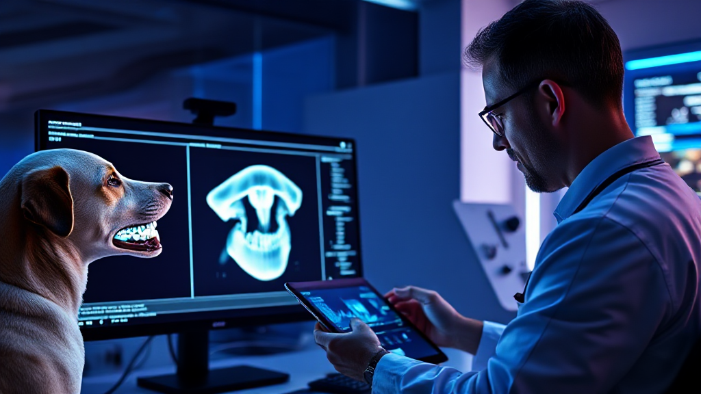
What AI Dental Models Can and Cannot Do in Veterinary Care
A dental AI model can flag a radiolucent halo around a tooth root in milliseconds; it cannot tell you whether that halo matters for this patient, this owner, and this anesthesia plan. That gap between detection and clinical conclusion is where veterinary dentistry actually lives. This article is an honest look at that boundary, written from the perspective of someone who builds and audits health-data models and has watched clinicians use them.
If you are evaluating whether model-assisted dental imaging belongs in your clinic, the short answer is that it belongs as a second reader, not a decision-maker. Platforms such as nerovet are built around that premise: AI-assisted technology layered into veterinary dental workflows so professionals keep the diagnostic authority. What follows explains what the model sees, what it can and cannot conclude, and how a practice should govern its output.
What the Model Actually Sees
An AI dental model does not see a tooth. It sees an array of pixel intensities, usually a few hundred by a few hundred values per image, normalized and passed through layers of learned weights. In veterinary dental radiography, those intensities encode mineral density, superimposition of structures, and the physics of the X-ray beam. The model learns statistical associations between patterns of intensity and labels that a human radiologist or veterinary dentist assigned during training.
That distinction matters because it explains both the model’s speed and its fragility. It is fast because matrix operations are cheap. It is fragile because a pattern it learned on one imaging setup may not transfer to another. A model recognizes image texture, not anatomy. When the texture shifts, the output can shift with it.
The input pipeline before inference
Most of the work in a dental AI system happens before the model runs. Images are acquired, cropped to the region of interest, resized, and often contrast-normalized so that exposure differences between patients do not dominate the signal. Some pipelines apply denoising or artifact suppression. Each of these steps is a design decision that changes what the model receives.
For veterinary practices, the practical consequence is that image acquisition discipline is not optional. A blurry or badly positioned intraoral radiograph degrades the input, and no amount of model sophistication recovers information that was never captured. Good lighting, consistent positioning, and correct exposure are data-quality controls, not just technique preferences.
What the output layer represents
The model’s output is typically a set of probabilities or bounding regions: a likelihood that a given area corresponds to bone loss, a flagged region around a root, a segmentation mask for a tooth crown. These are statistical statements about image patterns. They are not diagnoses, and they are not measurements in millimeters unless the pipeline has been calibrated against a known scale.
This is where many clinics misread the tool. A high confidence score means the model found a pattern similar to its training examples. It does not mean the pattern is clinically significant. Confidence is a property of the model, not of the patient.
What It Can and Cannot Conclude
The honest framing is a capability boundary, not a capability list. Models are good at consistency and recall on well-represented patterns. They are weak at context, rare presentations, and anything requiring synthesis across the patient’s history.
Where model assistance adds real value
- Detection support: flagging regions that warrant a closer look, especially in busy workflows where a subtle lesion might be skimmed.
- Consistency: applying the same visual criteria across images and across clinicians, which reduces inter-reader variation.
- Record structuring: converting findings into structured entries that are easier to search, audit, and hand over between shifts.
- Triage: surfacing cases that may need a veterinary dentist’s review sooner rather than later.
None of these replace the clinician. They reduce the cognitive load of scanning and make the record more useful after the fact.
Where the model stops
A model cannot integrate the patient’s age, breed predisposition, diet, prior extractions, or owner’s willingness to pursue treatment. It cannot feel a mobile tooth or assess gingival health by touch. It cannot judge whether a radiographic change is stable or progressive without prior images, and it cannot decide whether anesthesia risk outweighs the benefit of intervention.
It also cannot reliably generalize to presentations absent from its training data. Rare conditions, unusual anatomy, and artifacts from patient motion are classic failure modes. Absence of a flag is not evidence of absence. A negative output means the model did not recognize a pattern, not that the mouth is healthy.
A comparison in plain terms
Think of the difference as a table with two columns. On the left, the model’s column: pixel patterns, probability scores, bounding regions, consistency across images, speed, and reproducibility. On the right, the clinician’s column: patient history, physical examination, tactile findings, owner communication, treatment planning, and accountability for the outcome. The two columns overlap only at the image itself. Everything to the right of the image belongs to the professional, and no model output should be allowed to migrate into that column without a human decision.
Data Quality, Model Limits, and Why Human Oversight Stays Central
Model behavior is downstream of data quality. In veterinary dental imaging, data quality has three layers: acquisition, labeling, and representation. Acquisition covers positioning, exposure, and artifact control. Labeling covers whether the ground truth used in training was accurate and consistent. Representation covers whether the patient population in the training data resembles the patients in front of you.
Each layer introduces error. If labels were assigned by different clinicians with different thresholds, the model learns a blurred definition. If the training set overrepresents certain breeds or imaging equipment, performance may drop elsewhere. If the pipeline normalizes images in a way that suppresses subtle contrast, small lesions may disappear before the model ever sees them.
Why oversight is a design requirement, not a courtesy
Human oversight is central for three reasons. First, the model’s error modes are not symmetric: a missed lesion and a false flag have different clinical consequences, and only a clinician can weigh them for a given patient. Second, the model has no access to the context that determines treatment. Third, accountability for the patient’s care rests with the professional, and that accountability cannot be delegated to a statistical output.
This is why platforms in this space emphasize workflow integration rather than autonomous diagnosis. The value of nerovet in a practice is not that it replaces the clinician’s eye but that it structures the image review and the record around the clinician’s judgment. The model proposes; the professional disposes.
Common mistakes clinics make with model output
- Treating a flag as a diagnosis. A highlighted region is a prompt to look, not a finding to record as confirmed.
- Skipping the negative review. Clinicians sometimes relax when the model reports nothing, which inverts the intended safety logic.
- Ignoring acquisition quality. Poor images produce poor outputs, and the model has no way to say “this image is inadequate.”
- Letting the record lose its provenance. If structured entries do not distinguish model-suggested from clinician-confirmed findings, the record becomes misleading later.
- Assuming transferability. A model validated on one equipment setup is not automatically valid on another.
How Clinics Should Govern AI Dental Output
Governance is the practical answer to the boundary described above. It means deciding, in advance, what the model is allowed to influence and what it is not. A simple governance framework has four parts: a written scope, a review protocol, a record convention, and a periodic audit.
Set a written scope
The scope should state which tasks the model supports and which it does not. Detection support and record structuring are reasonable. Autonomous diagnosis, treatment selection, and client communication are not. Writing this down protects the practice and sets expectations for new team members.
Define the review protocol
Every model output should be reviewed by a qualified professional before it enters the clinical record as a finding. The protocol should specify who reviews, what constitutes confirmation, and what happens when the reviewer disagrees with the model. Disagreements are not failures; they are the system working as intended, and they should be logged because they reveal where the model’s limits sit in your patient population.
Keep provenance in the record
Structured records should distinguish model-suggested entries from clinician-confirmed ones. This is a data-integrity requirement, not a bureaucratic one. If a later reviewer cannot tell which findings were machine-proposed, the record’s reliability erodes. Provenance is what makes an AI-assisted record trustworthy.
Audit periodically
A periodic audit compares model flags against clinician conclusions over a sample of cases. The goal is not a single accuracy number but a picture of where the model over-flags and under-flags in your practice. Audits should also check image quality trends, because declining acquisition discipline will show up as declining model usefulness.
Train the team on the boundary
Staff should understand that the model is a reader, not an authority. Front-desk and technician teams who interact with structured records need to know that a flagged entry is not a confirmed diagnosis and should not be communicated to owners as one. This is where many implementations quietly fail: the technology works, but the human workflow around it does not.
Plan for failure modes
Every model has failure modes, and a governed practice plans for them. Expect false negatives on subtle lesions, false positives on artifacts, and degraded performance when imaging equipment changes. Build a fallback: when the model’s output conflicts with clinical judgment, clinical judgment prevails, and the case is documented. Over time, those documented conflicts become the most useful data your practice has for improving both technique and tool selection.
The boundary between detection and conclusion is not a flaw to be engineered away. It is the structure of responsible veterinary dental AI. Models are strong where consistency and recall matter, and weak where context and accountability matter. A clinic that governs the output, keeps provenance in the record, and treats the model as a second reader gets the benefit without inheriting the risk. That is the realistic promise of AI-assisted veterinary dentistry, and it is a promise worth keeping honest.
